Kiel Cardio Database
|
All measurements were approved by the ethics committee of Kiel University (File number: A 117/20) and conducted in accordance with the Declaration of Helsinki. |
 The "KCD" corpus (Kiel Cardio Database) currently consists of one-minute 8-lead MCG (magnetocardiographic) measurements from seven subjects. For each subject, 25 subsequent measurements were made with the sensor array, consisting of four QuSpin QZFMs, at different positions for each trial. Therefore, 200 MCG measurements are available for each subject. The database includes the raw and preprocessed recordings.
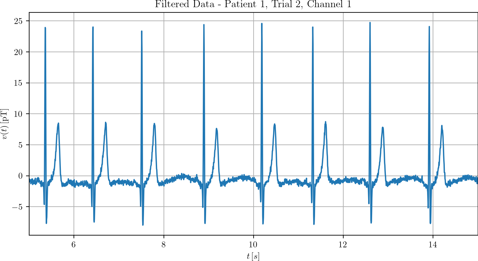
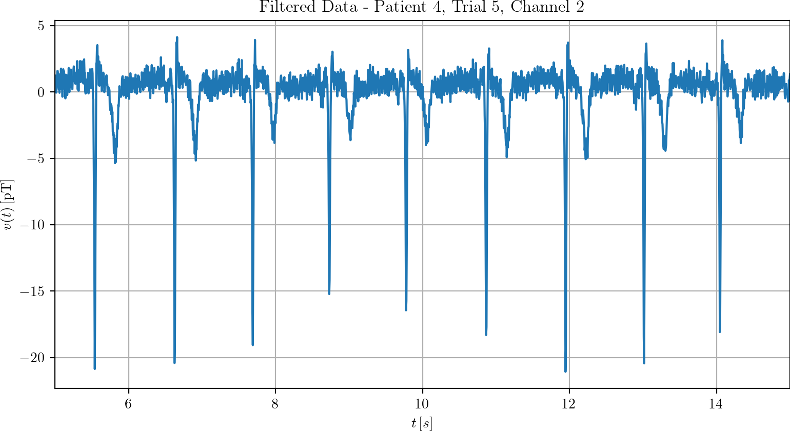
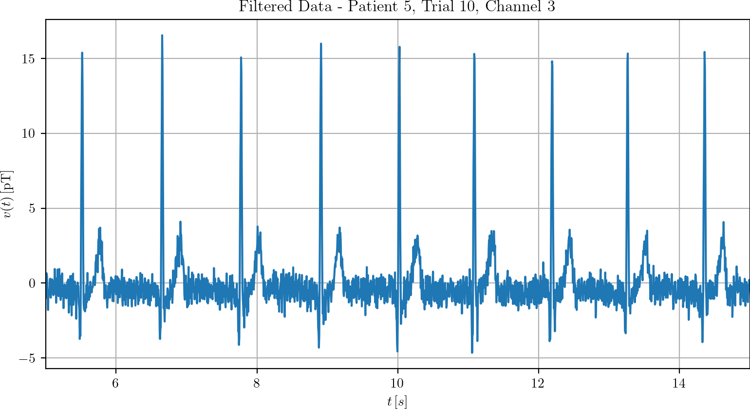
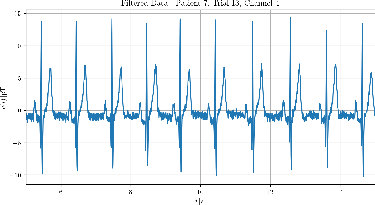
Examples
Below you can find examples of the preprocessed recordings.
|
|
|
|
|
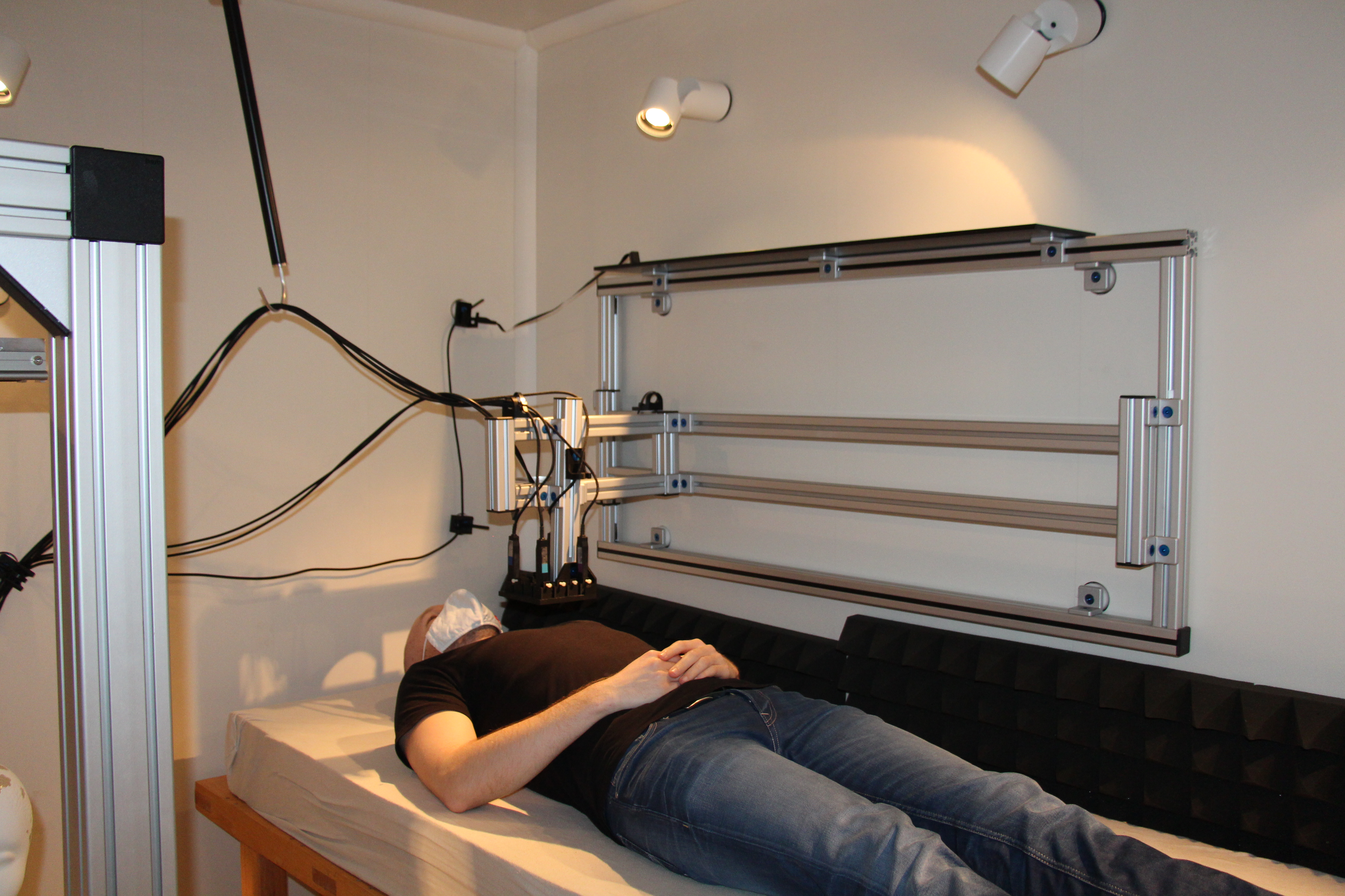
Measurement Setup
Coordinate Systems
|
The second coordinate system refers to the sensor array. The direction of the axes is the same as for the first coordinate system and the origin is at the center of the tip of sensor 0. |
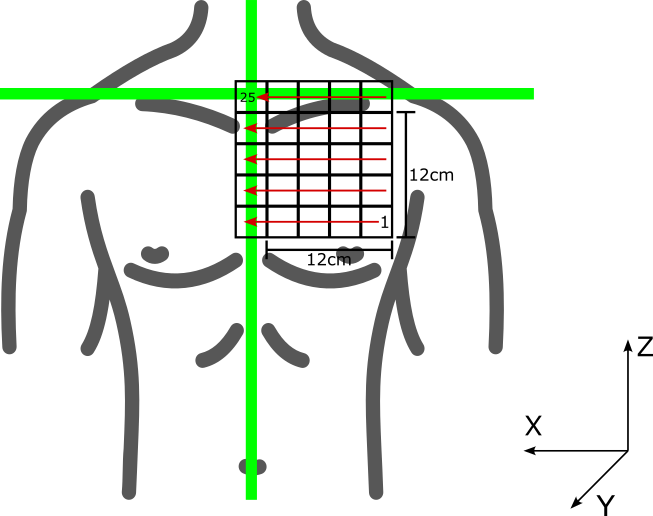 Two coordinate systems are used for this database. The first, global, coordinate system refers to the subject. The x-axis runs slightly above the shoulders from left to right. The y-axis is from the subject's back to the front. The z-axis is aligned from the subject's spine and points from feet to head. The point (0,0,0) is therefore located on the bed at the intersection between the patient's spine and an a orthogonal line touching the top of the shoulders.
Sensors
|
|
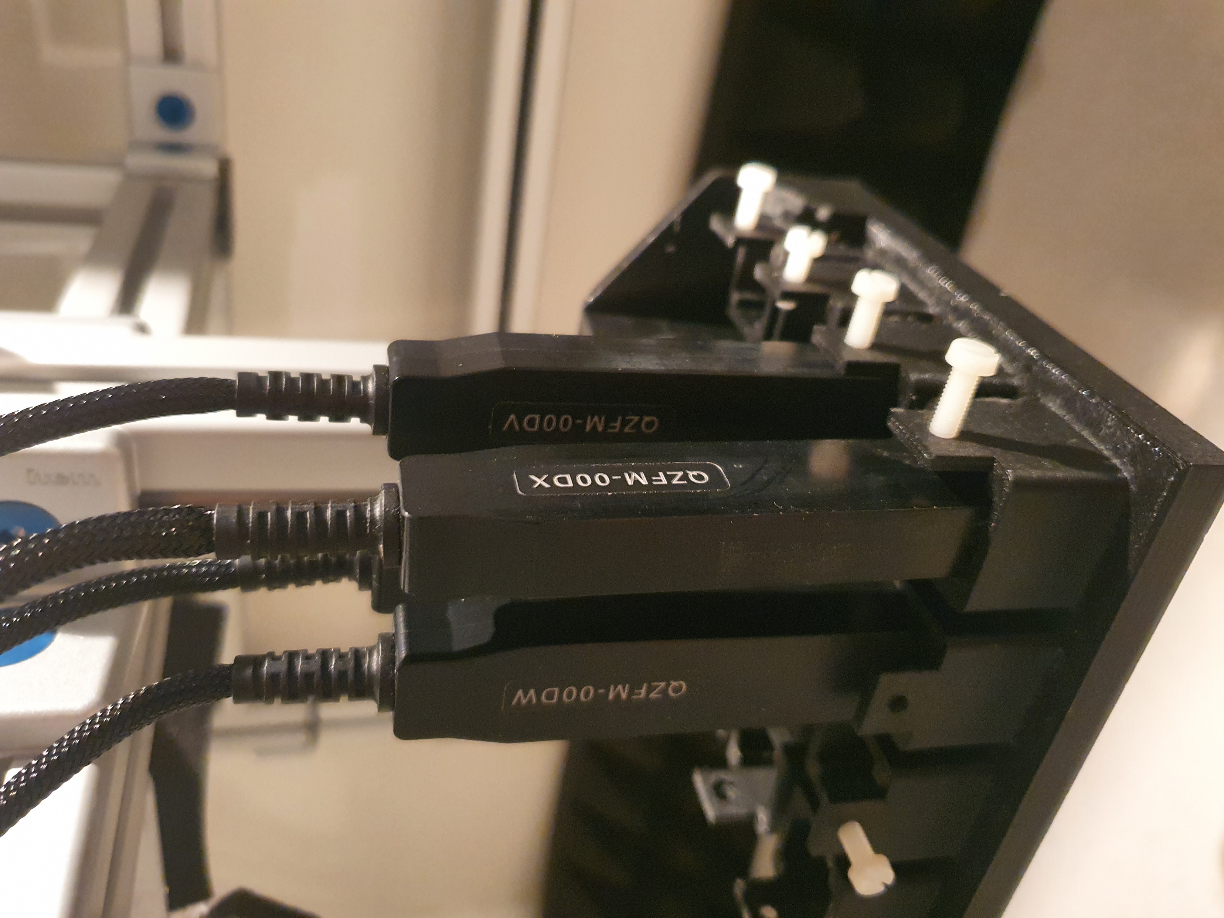 The sensors used are four QuSpin QZFM Gen 1 in two-axis mode. They are referred to below as sensor 0, sensor 1, sensor 2, and sensor 3. The sequence and measuring directions of the sensors are listed in the table below.
Location
All measurements were performed in the magnetically shielded chamber at the Faculty of Engineering of the Christian-Albrechts-Universität zu Kiel.
Software
The real-time software KiRAT, which was developed by the Digital Signal Processing and System Theory (DSS) group, was used to drive and read out the sensors.
Positions
|
The positions of the sensors in relation to the center of the sensor array are listed in the table below.
The position of the sensor array in regards to the subject origin in trial 1 is (-11 cm, 17 cm, -14 cm). |
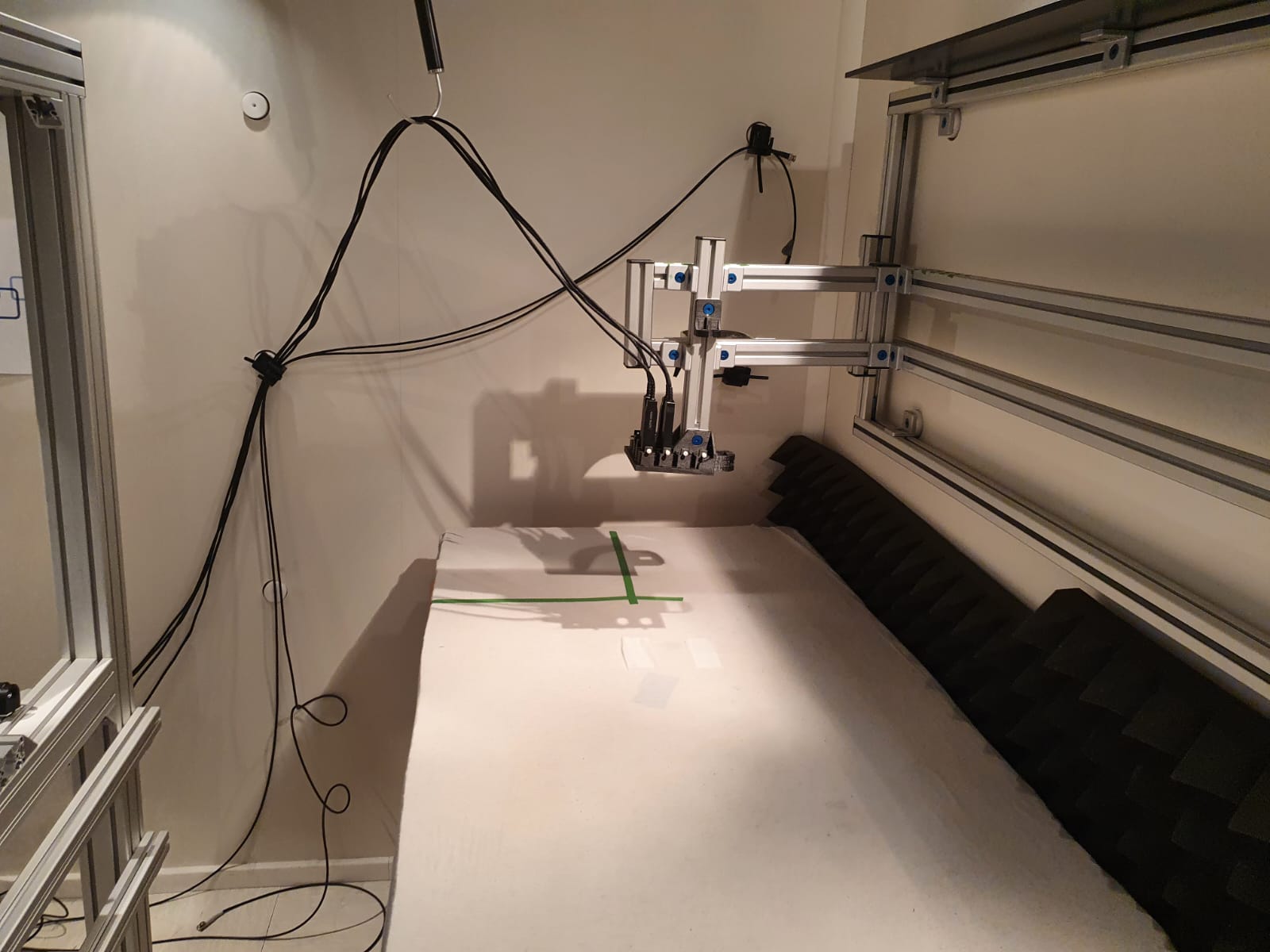 Below is information on the sensor positions during the various trials. All four sensors were placed in a fixture, which was then moved between trials. To keep the information as clear as possible, it was divided into three parts. The relative position of the sensors in the holder/sensor array, the "normal" position of the sensor array during the first trial, and the position offset for the following 24 trials. All positions are given in centimeters.
 The y-offset is zero for all trials. Below you can see an overview with the x-offset in the column and the z-offset in the rows (as in the figure above).
| Offsets | 12 cm | 9 cm | 6 cm | 3 cm | 0 cm |
|---|---|---|---|---|---|
| 12 cm | 25 | 24 | 23 | 22 | 21 |
| 9 cm | 20 | 19 | 18 | 17 | 16 |
| 6 cm | 15 | 14 | 13 | 12 | 11 |
| 3 cm | 10 | 9 | 8 | 7 | 6 |
| 0 cm | 5 | 4 | 3 | 2 | 1 |
Outlook
While the corpus currently consists only of the raw MCG data, we plan to expand it in the future. Some features you can expect in the future are:
- 144 channel ECG recordings
- Segmentation data for the ECG and MCG signals (P wave, QRS complex, T wave).
- (segmented) MRI/CT scans of the subjects' torsos
- Heart surface potential of selected subjects (only those who have to undergo the procedure anyway due to medical conditions)